Radiation and Medical X-rays
Radiation Facts
- As with any medical test that uses radiation, what is learned from an x-ray procedure should provide a benefit that outweighs the radiation risk.
From broken bones to life threatening illnesses, x-rays help doctors find and treat medical conditions. Specially trained doctors can read these images to diagnose medical conditions or injuries. Different imaging procedures expose patients to different amounts of radiation. For the average American, medical x-rays are their single largest source of man-made radiation exposure.
About Radiation and Medical X-rays

Medical x-rays are used to help doctors see what is happening inside the body. X-rays pass through objects, including internal organs, body tissue and clothing. The x-rays project a picture onto film or send a digital image to a computer. Bones appear white on x-ray images because denser objects absorb more radiation. Less dense parts of the body, such as skin and muscles, remain dark on x-ray images because the radiation passes through them. A medical x-ray produces a picture that can help find broken bones, tumors and foreign objects in the body. X-rays are also used in other types of examinations and procedures, including CT scans, mammograms and fluoroscopy. Medical x-rays, dental x-rays, and mammograms use relatively low amounts of radiation. CT scans and fluoroscopic procedures result in higher radiation doses due to the need for multiple images and/or a longer exposure time. However, medical imaging is a very powerful and valuable technique that can provide important and lifesaving information.
Dental X-rays
Dental x-rays are pictures of your teeth from the crown to the roots. They allow the dentist to see inside and between your teeth and check the overall condition of the bones of your jaw and face. During a dental x-ray, radiation passes through your cheek and gums and creates an image using the special x-ray film clamped between your teeth. Some x-ray machines create a digital image instead of using film. Conventional dental x-rays use a small amount of radiation to take the pictures. More detailed images may be needed in certain circumstances, such as planning for orthodontics or dental implants. In these cases, cone beam computed tomography may be used, which results in higher doses than other dental examinations (for more information, see CT Scans below).
Mammography
A mammogram is an x-ray picture of breast tissue that is used to detect breast cancer. There are two kinds of mammograms, screening and diagnostic. Screening mammograms are used to check healthy women with no signs of disease. Screening mammograms use very small doses of x-ray radiation. Diagnostic mammograms are helpful when there are some symptoms of breast cancer. Diagnostic mammograms often include more than a single x-ray. X-rays from multiple angles are needed, therefore patients who have diagnostic mammograms are exposed to more radiation. The risk of not having a needed mammogram is greater than the risks from radiation. Mammograms are a valuable tool in the early detection of breast cancer.
CT Scans
Computed tomography scans (also known as CT scans, CAT scans, or computed axial tomography scans) are x-ray procedures that create cross-sectional views and three-dimensional images of a patient's internal organs. When a person has a CT scan, many x-rays are taken at nearly the same time. CT scans create very clear images of internal organs. These detailed images help doctors diagnose problems inside the body, such as tumors or organ damage. CT scans can also provide surgeons a map of the inside of the patient that they can follow when operating. When a person has a CT scan, they are being exposed to up to several hundred times more radiation than a conventional x-ray. Like any medical test, the beneficial information gained from a CT scan should outweigh the risk of radiation exposure from the test performed. When needed, CT scans are a very powerful and valuable technique that can provide important and lifesaving information.
Fluoroscopy
Fluoroscopy uses x-rays to show movement in real-time. It can show the movement of a body part, like the beating of a heart, or the path taken by a medical instrument or dye (contrast agent) as it moves through the body. Unlike conventional x-rays, fluoroscopy uses an intermittent, pulsed x-ray beam that is passed through the body. The images are sent to a monitor where doctors can see the body part and its motion in real-time. The total radiation exposure from x-rays depends on the length of time of the fluoroscopic procedure and how often the x-ray beam is used. Fluoroscopy is used in many types of examinations and procedures, including:
- Viewing movement of materials through the stomach and intestines
- Directing the placement of a catheter during heart surgery
- Visualizing blood flow to organs
- Helping doctors properly set broken bones
What You Can Do
- Ask your doctor how an x-ray will help. How will an x-ray, CT scan, or fluoroscopy procedure help find out what is wrong or determine treatment? Ask if there are other test that might use less or no radiation.
- Don't refuse an x-ray that is needed. If your healthcare provider explains why an x-ray is medically needed, then do not refuse the test. The risk of not having a needed x-ray is greater than the risk from radiation.
- Don't insist on an x-ray. If your doctor tells you that an x-ray is not needed, then do not demand one.
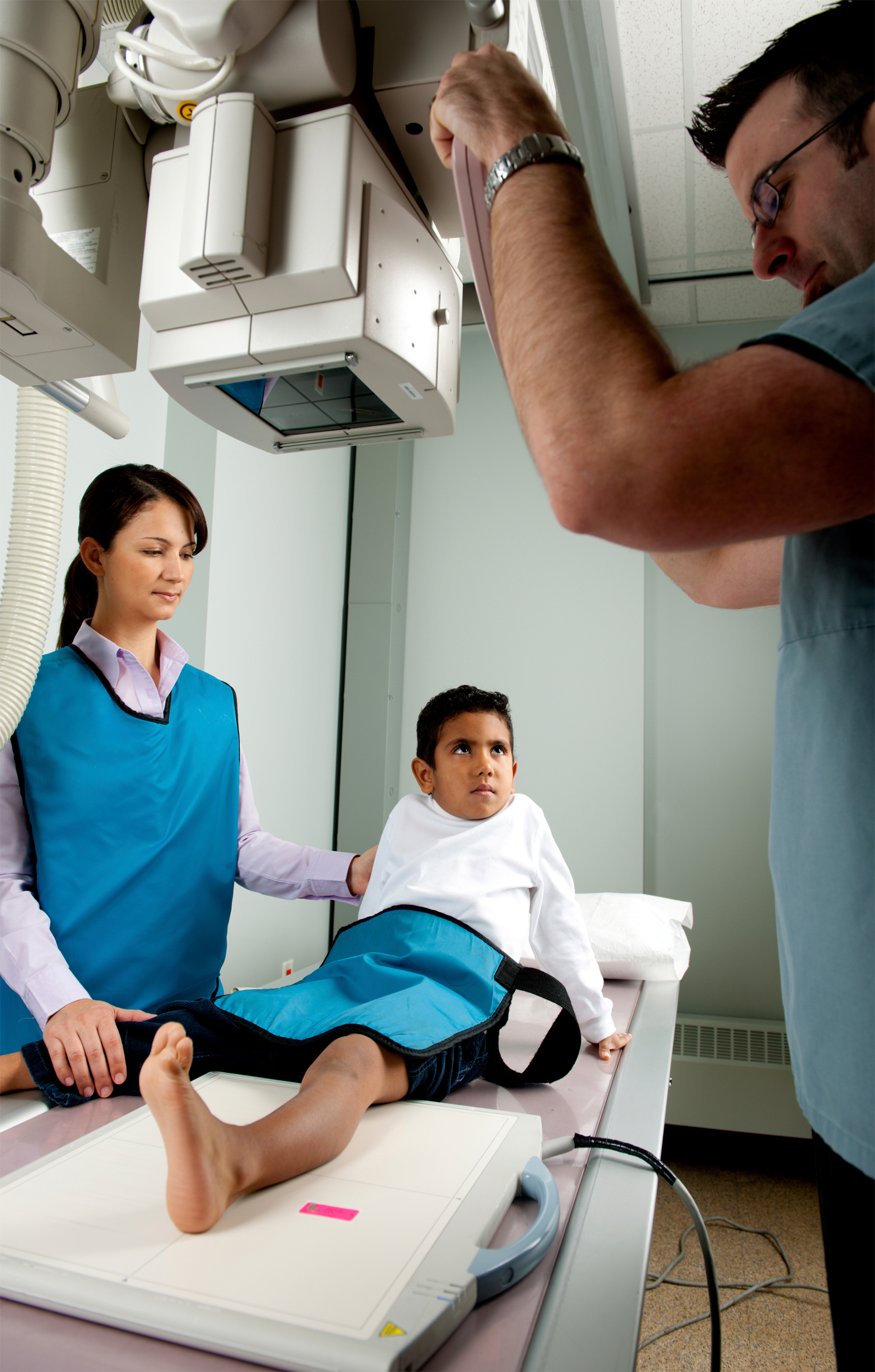
- Tell the x-ray technologist if you are, or might be, pregnant. This will allow the doctor or technician to take steps to limit radiation to the fetus.
- Ask if a protective shield can be used. If you or your children are getting an x-ray, ask whether a lead apron or other shield is appropriate.
- Ask your dentist about the type of x-ray film used. Ask if your dentist uses the faster E or F speed film for x-rays. It costs about the same as the conventional D speed film and offers similar quality images with a lower radiation dose. Many dentists now use digital x-ray imaging instead of film, which further reduces radiation dose as long as image retakes are minimized.
- Know your x-ray history. Keep a list of your imaging records, including dental x-rays. When an x-ray is taken, fill out the card with the date and type of exam, referring doctor, and the name and address of where the images are kept. Maintaining and referring to this record can help you avoid unnecessary duplicate x-rays. Keep a record card for everyone in your family.